Statistical Modelling
Acetabular Reconstruction
Planning hip reconstruction in patients with large acetabular defects is difficult due to the lack of anatomical landmarks, as a result of the deformed anatomy and contralateral hip being often replaced.
Statistical Shape Models (SSMs) are mathematical models that can be used to successfully reconstruct the absent bony landmarks of diseased anatomies regardless of the severity of the defects.
SSMs are important tools to estimate the original position of the hip joint centre prior to the development of the bony defect, and it is a valid starting point for engineers to design customised implants for the treatment of patients with large acetabular defects.
Femoral Reconstruction
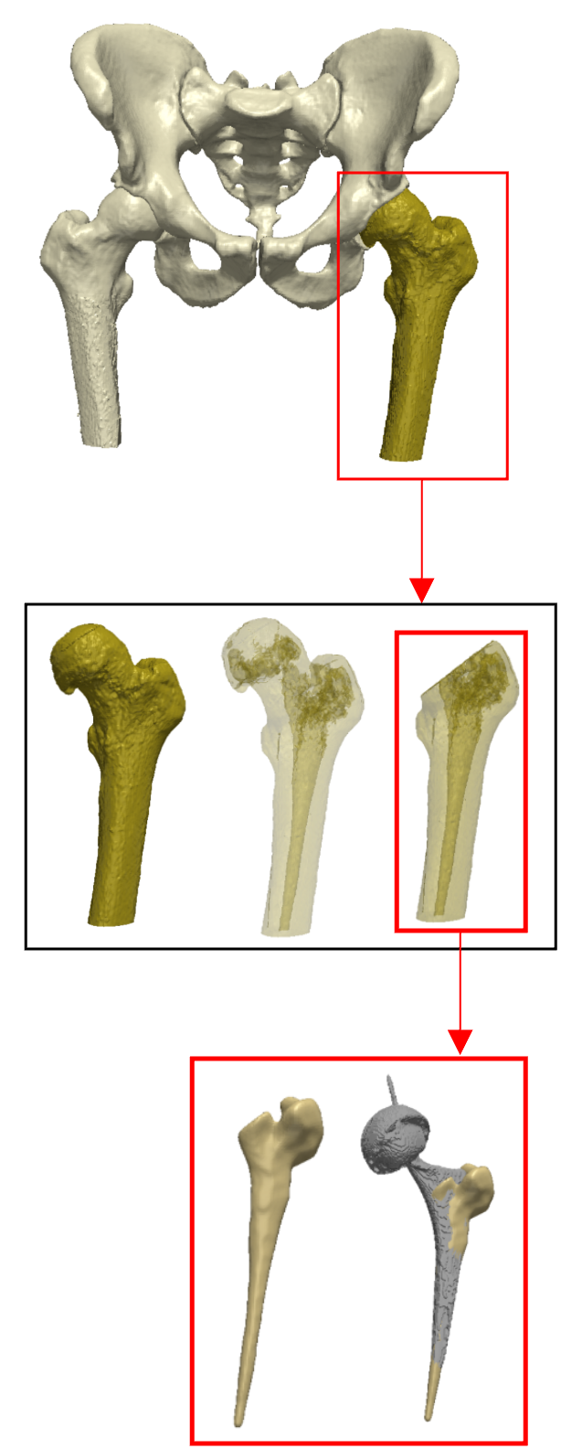
SSM may also be applied to the other side of the hip-joint; the femur. In uncemented hip replacement the orientation of the femoral stem is largely guided by the intramedullary canal of the proximal femur. As a result, the surgeon has limited intraoperative control over the achieved position of the implant.
At present, the femoral component position cannot be accurately planned due to a lack of understanding of the shape of the femoral canal. SSM offers a useful tool to identify the key distinguishing anatomical features of this femoral region.
Our Publications
Computational modelling of acetabular morphology and its implications for cup positioning
Computational modelling of acetabular morphology and its implications for cup positioning
Statistical Shape Modelling of the Large Acetabular Defect in Hip Revision Surgery
Statistical Shape Modelling of the Large Acetabular Defect in Hip Revision Surgery
Understanding the Variability of the Proximal Femoral Canal: A Computational Modeling Study
Understanding the Variability of the Proximal Femoral Canal: A Computational Modeling Study
Hip reconstruction in patients with large acetabular defects is challenging due to their lack of anatomical landmarks, the result of the deformed anatomy and the contralateral hip being often replaced. In addition, failed implants can create metal artefacts that obscure bony readings from CT scans, making 3D reconstruction challenging. The SSM workflow in this case study overcomes these limitations by successfully reconstructing the absent bony landmarks of diseased anatomies, regardless of the severity of the defects.

